抗体来源(Source)
Biotinylated Monoclonal Anti-TNF-alpha Antibody, human IgG1 (16H5) is a chimeric monoclonal antibody recombinantly expressed from HEK293, which combines the variable region of a mouse monoclonal antibody with Human constant domain.
克隆号(Clone)
16H5
亚型(Isotype)
Human IgG1 | Human Kappa
偶联(Conjugate)
Biotin
抗体类型(Antibody Type)
Recombinant Monoclonal
种属反应性(Reactivity)
Human
免疫原(Immunogen)
Human TNF-alpha Protein is expressed from human 293 cells.
特异性(Specificity)
This product is a specific antibody specifically reacts with TNF-alpha.
应用(Application)
| Application | Recommended Usage |
| ELISA | 0.2-50 ng/mL |
纯度(Purity)
>95% as determined by SDS-PAGE.
>90% as determined by SEC-MALS.
纯化(Purification)
Protein A purified / Protein G purified
制剂(Formulation)
Lyophilized from 0.22 μm filtered solution in PBS, pH7.4 with trehalose as protectant.
Contact us for customized product form or formulation.
重构方法(Reconstitution)
Please see Certificate of Analysis for specific instructions.
For best performance, we strongly recommend you to follow the reconstitution protocol provided in the CoA.
存储(Storage)
For long term storage, the product should be stored at lyophilized state at -20°C or lower.
Please avoid repeated freeze-thaw cycles.
This product is stable after storage at:
- -20°C to -70°C for 12 months in lyophilized state;
- -70°C for 3 months under sterile conditions after reconstitution.
电泳(SDS-PAGE)
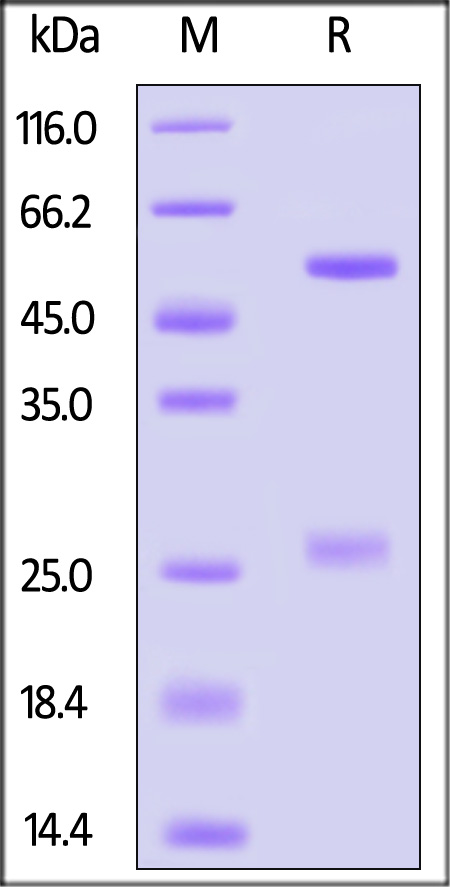
Biotinylated Monoclonal Anti-TNF-alpha Antibody, human IgG1 (16H5) on SDS-PAGE under reducing (R) condition. The gel was stained with Coomassie Blue. The purity of the protein is greater than 95%.
SEC-MALS
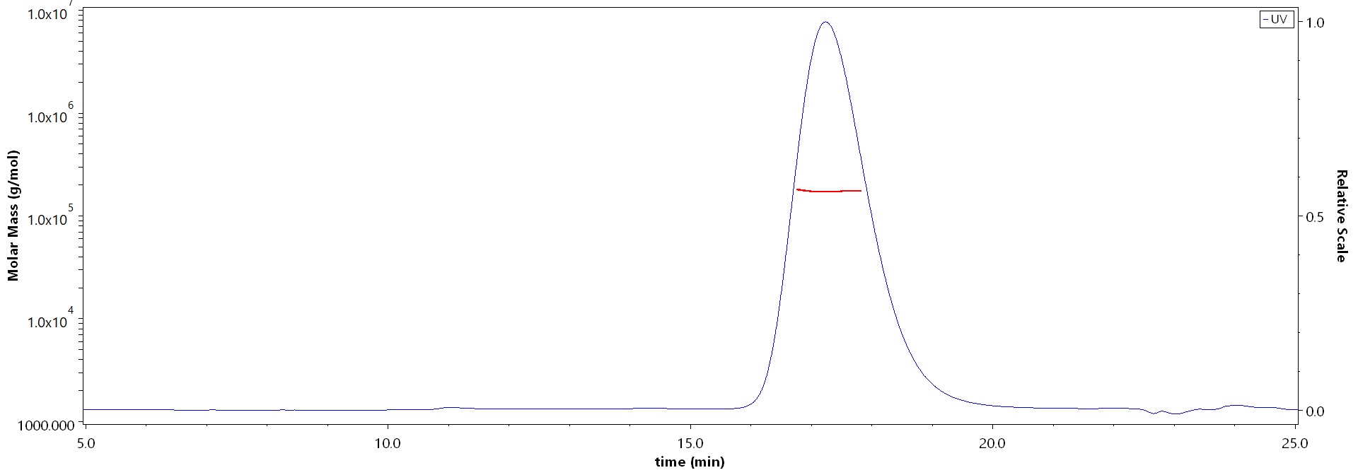
The purity of Biotinylated Monoclonal Anti-TNF-alpha Antibody, human IgG1 (16H5) (Cat. No. TNA-BLM494) is more than 90% and the molecular weight of this protein is around 135-175 kDa verified by SEC-MALS.
Report
活性(Bioactivity)-ELISA
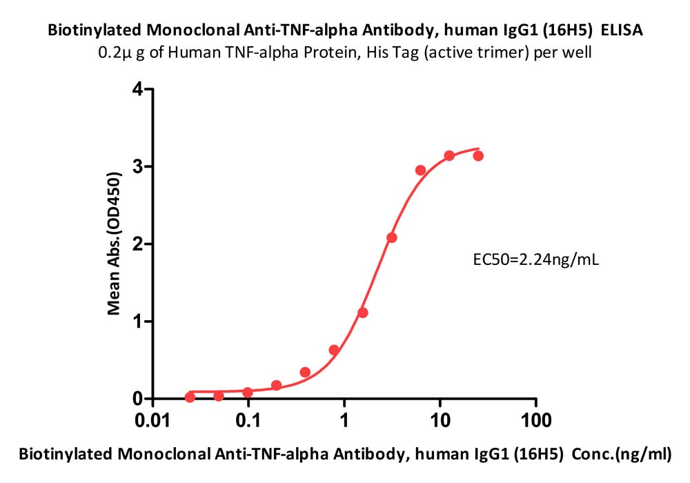
Immobilized Human TNF-alpha Protein, His Tag (Cat. No. TNA-H5228) at 2 μg/mL (100 μL/well) can bind Biotinylated Monoclonal Anti-TNF-alpha Antibody, human IgG1 (16H5) (Cat. No. TNA-BLM494) with a linear range of 0.20-3.13 ng/mL (QC tested).
Protocol
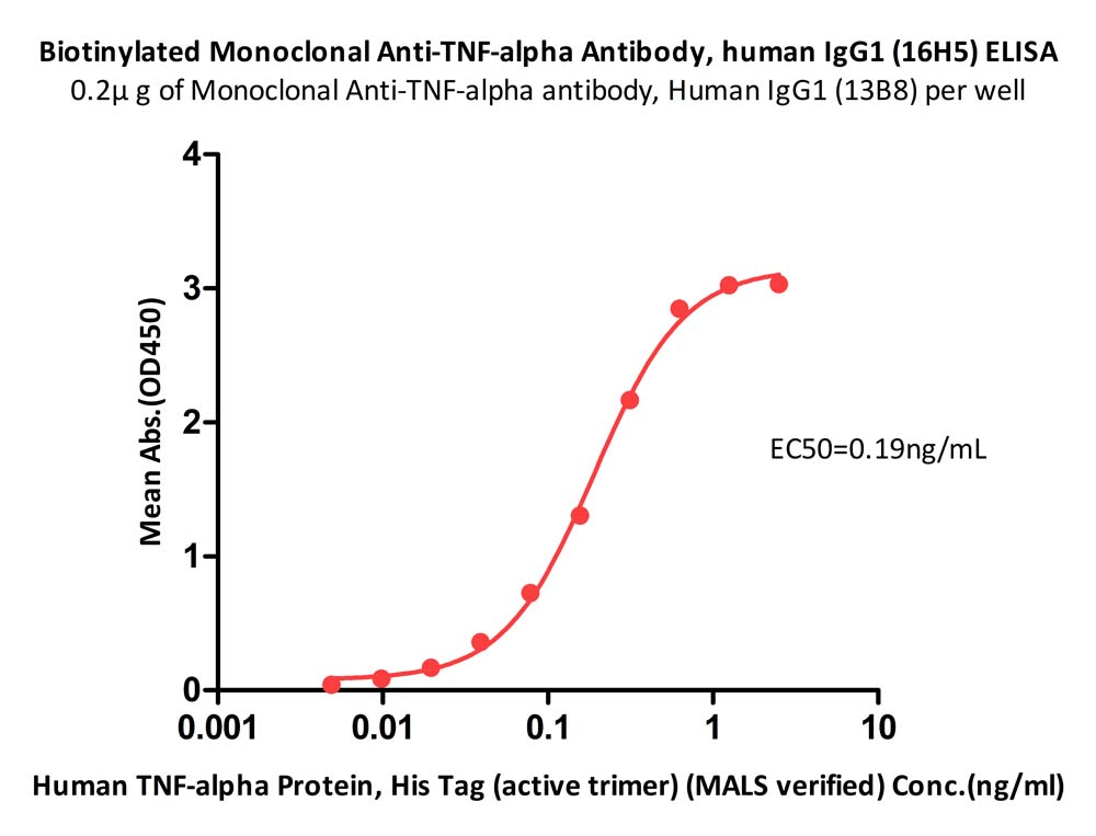
Immobilized Monoclonal Anti-TNF-alpha Antibody, Human IgG1 (13B8) (Cat. No. TNA-AM493) at 2 μg/mL, add increasing concentrations of Human TNF-alpha Protein, His Tag (Cat. No. TNA-H5228), and then add Biotinylated Monoclonal Anti-TNF-alpha Antibody, human IgG1 (16H5) (Cat. No. TNA-BLM494) at 0.25 μg/mL. Detection was performed using HRP-conjugated streptavidin with sensitivity of 0.02 ng/mL (Routinely tested).
Protocol
活性(Bioactivity)-SPR

Biotinylated Monoclonal Anti-TNF-alpha Antibody, human IgG1 (16H5) (Cat. No. TNA-BLM494) captured on CM5 chip via Anti-human IgG Fc antibodies surface can bind Human TNF-alpha, His Tag (Cat. No. TNA-H5228) with an affinity constant of 75.9 pM as determined in a SPR assay (Biacore 8K) (Routinely tested).
Protocol
活性(Bioactivity)-BLI
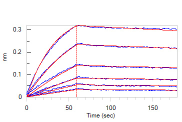
Loaded Biotinylated Monoclonal Anti-TNF-alpha Antibody, human IgG1 (16H5) (Cat. No. TNA-BLM494) on Protein A Biosensor, can bind Human TNF-alpha, His Tag (Cat. No. TNA-H5228) with an affinity constant of 2.13 nM as determined in BLI assay (ForteBio Octet Red96e) (Routinely tested).
Protocol
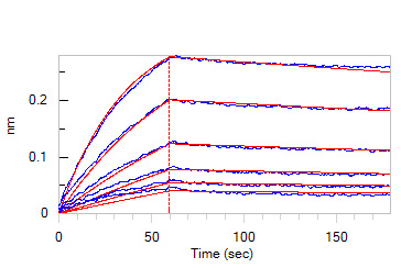
Loaded Biotinylated Monoclonal Anti-TNF-alpha Antibody, human IgG1 (16H5) (Cat. No. TNA-BLM494) on Protein A Biosensor, can bind Human TNF-alpha, premium grade (Cat. No. TNA-H4211) with an affinity constant of 3.16 nM as determined in BLI assay (ForteBio Octet Red96e) (Routinely tested).
Protocol
背景(Background)
Tumor necrosis factor alpha (TNFα) is a cytokine produced primarily by monocytes and macrophages. It is found in synovial cells and macrophages in the tissues.The primary role of TNFα is in the regulation of immune cells. TNFα is able to induce apoptotic cell death, to induce inflammation, and to inhibit tumorigenesis and viral replication. Dysregulation of TNFα production has been implicated in a variety of human diseases, including major depression, Alzheimer's disease and cancer. Recombinant TNFα is used as an immunostimulant under the INN tasonermin. TNFα can be produced ectopically in the setting of malignancy and parallels parathyroid hormone both in causing secondary hypercalcemia and in the cancers with which excessive production is associated.


 膜杰作
膜杰作 Star Staining


